KG MediChat
|
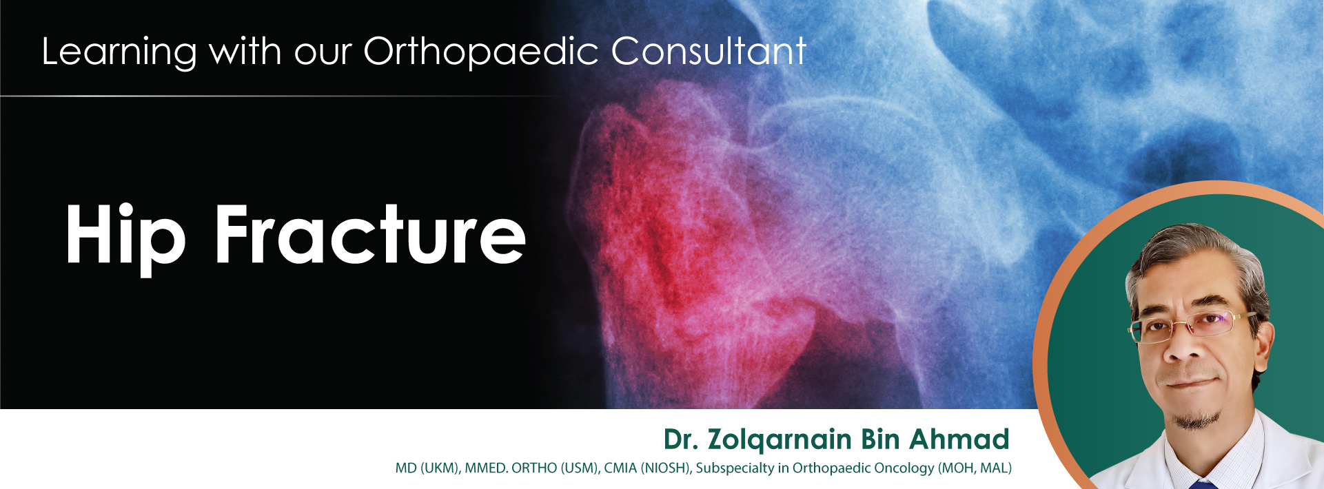
| Hip Fracture in Elderly
|
Dr. Zolqarnain Bin Ahmad
|
|
| A hip fracture may result in a loss of physical function, decreased social engagement, increased dependence, and worse quality of life. It can be a life-threatening illness for elderly patient. Death after a hip fracture may also be related to additional complications of the fracture, such as infections, internal bleeding, stroke, or heart failure.
|
|
|

|
|
Reasons/ Causes/ Risk Factors & ComplicationHip Fracture in Elderly
|
|
|
|
- Many hip fractures occur from injures such as a fall.
- Osteoporosis is one condition that causes hip fractures.
- Elderly with weak bones, a hip fracture can occur simply by standing on the leg or sitting down.
- Age is a key risk factor, with hip fractures more likely to occur in those aged 65 or older.
- 1 in 3 adults aged 50 and over dies within 12 months of suffering a hip fracture.
- Older adults have a 5 - 8 times higher risk of dying within the first 3 months of a hip fracture compared to those without a hip fracture.
- Research shown around 30% of people with hip fractures have had a prior fracture; this is known as the “fracture cascade”.
Some of the more common problems that a hip fracture can increase the likelihood of include:
- Pneumonia
- Bed sores (pressure ulcers)
- Deep Vein Thrombosis (DVT) – blood clots in the large veins of the leg
- Mental confusion
|
|
|
|
|
|
Treatment Option & Doctor Perception
|
|
|
|
| The goal of any hip fracture surgery is to hold the broken bones securely in position, allowing the patient to get out of bed as soon as possible. Hip fractures in the elderly are usually treated with surgery to fix the fractured bones after proper assessment, it is normally done within 24 hours of admission to the hospital.
|
|
|
| Most hip fractures are treated in one of three ways: with metal pins, a metal plate and screws, or with artificial replacement of the broken femoral head. Upon consulting, doctor will evaluate the risk and urgency of the patient’s condition.
|
|
|
| Some cases of hip fractures would actually heal without surgery involved, but it will take 8 – 12 weeks for patient to recover. However, placing an elderly person in bed for a long period of time may results in greater risk of creating serious complications than the surgery. This is the reason surgery is recommended to nearly all patients with fractured hips.
|
|
|
|

|
|
Prevention & Rehabilitation
|
|
|
|
How do we prevent hip fractures and taking care of your bones?
- Do strength and balance exercises to strengthen your muscle and bones
- Ensure your home is safe for elderly, getting eye exam to protect your vision
- Consult a doctor to evaluate your risk and review medicines
- Get an Osteoporosis screening / bone density test
|
|
|
|

|
|
Rehabilitation After a Hip Fracture
|
|
|
|
| Most patients are able to start weight bearing right away after surgery. Depending on the severity of the fracture, patients may only be able to place partial weight down right away. To speed up the recovery, therapeutic rehabilitation will be involved. Physical therapy may be needed for patients who have problems walking.
|
|
|
|
|
| Enterovirus Infection
|
Dr. Choong Choun Seng
|
.jpg)
|
| Malaysia is like summer all the year round. Major and minor epidemiological cases of enteroviruses occur every month. With the peak from March to June, the symptoms are mostly manifested by Herpangina and Hand, Foot, and Mouth Disease (HFMD). Most of the enterovirus infections are mild and will recover after a certain course of disease. This type of virus infection is mainly caused by the Coxsackie virus. The few types that can cause severe illness or even death are mainly enterovirus type 71.
|
|
|
Clinical manifestations:
|

|
| Herpangina
|
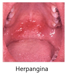
- Mainly caused by the Coxsackie A virus.
- Characteristics: Sudden on set of high fever, sore throat and vomiting, blisters or ulcers in the pharynx, inability to eat, and some require hospitalization and intravenous therapy. The course of the disease is 4-6 days.
- Mainly affects children 1-7 years old. The main route of infection is Fecal-Oral or respiratory tract.
- Key prevention method: Wash your hands frequently.
|
|
|
|
|
| Hand, Foot, and Mouth Disease (HFMD)
|
- The most common causes of the virus are Coxsackie A, B virus and enterovirus type 71.
- Characteristics: Fever, small blisters
- Mainly distributed in oral mucosa and tongue, followed by soft palate, teeth, and lips. The blisters over the limbs consist of the palms and soles of the feet or in between the fingers and toes. Occasionally, there are small blisters on the butt, knees, and elbows. Some blisters are itchy, and the general course of the disease is about seven days.
|

|
|
|
|
| Infant acute Myocarditis
|
- Mostly caused by Coxsackie virus type B and Enterovirus Type 71
- Characteristics: Sudden breathing difficulties, pallor, fever, vomiting, followed by rapid heartbeat, heart failure and shock, and even death.
- If your children have the symptoms of poor activity, continuous vomiting, rapid heartbeat (Tachycardia), and Myoclonic Jerk, please send them to the hospital immediately.
|
|
|
|
|
|
|
|
|
|
|
|
|
What you need to know about dengue?
|
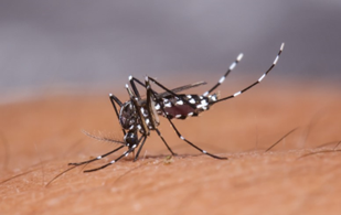
Dengue is a very common disease in Malaysia that occurs as a result of the bite of a disease carrying mosquito called Aedes. This mosquito typically comes out to feed at dawn and dusk.
After being infected, the disease symptoms typically range from dengue fever (DF) that heals by itself, to dengue haemorrhagic fever (DHF) and DHF with shock syndrome (this means one’ll be very sick and MUST be admitted, as this is potential life-threatening). The risk of severe disease is much higher if one gets dengue a second time than the first.
|

|
|

|
| Classical DF is a newly occurring fever accompanied by headache, pain behind one’s eye balls & marked muscle and bone pains. Fever lasts typically for 5 to 7 days, during which at this time one’s platelet (the component of the blood that’s used for stopping a bleed from a wound) will be low & with or without the typical rash (what doctors call “Isles of white in a sea of red”).
|

|

|

|
| DHF is the most serious manifestation of dengue infection and plasma leakage (fluid that escapes from the cells into other bodily spaces such as lungs and stomach) is the most life-threatening feature of DHF. This usually occurs over a period of 24 to 48 hours AFTER fever has subsided (Yes, NOT DURING fever). Haemorrhagic manifestation in DHF can range from spontaneous petechiae (like the red rash in the picture) or profuse bleeding (e.g., like throwing up blood).
|

|
The diagnosis of DF is based on signs and symptoms in individuals exposed to dengue (in Malaysia, while some places are labelled as “black dengue areas”, literally EVERYWHERE is dengue prevalent). A simple blood test at a hospital (and certain clinics) will be able to help doctors confirm DF.
Typically, we advise the public to come see a doctor the moment you have a fever lasting at or beyond 3 days. But we strongly advise medical attention if one has any dengue warning signs (regardless of number of fever days), which are:
|

|
- Abdominal pain
- Persistent vomiting
- Any bleeding (e.g., gum bleed, bruises, or vomiting blood)
- Restlessness or excessive lethargy
- Difficulty breathing, leg/hand/facial swelling or severe loss of appetite (due to fluid accumulation in the designated parts of the body)
|
|
|
|
|
|
|
|
| Prenatal Testing for Down Syndrome
|
Dr. Hoo Woon Ping
|
|
Prenatal Testing For Down Syndrome
|
| What is Down Syndrome (also called Trisomy 21 or T21)?
|
| Down syndrome is a condition in which a person carries an extra copy of chromosome 21. This extra copy of chromosome 21 changes how the baby’s body and brain develop and can result in learning difficulties, heart conditions or problems with digestive tract, hearing, and vision. Sometimes this can be serious, but many can be treated.
|
|
|
| Who should have the screening test for Down Syndrome?
|
| All women, regardless of age, should be offered screening tests to assess your chances of carrying a baby with Down Syndrome. You do not need to have a test – it is your choice. Some people want to find out the chance of their baby having Down syndrome, and some do not.
|
|
|
| What do screening test for Down Syndrome involve?
|
- First trimester screening test combines a harmless blood test with an ultrasound scan to measure a specific area on the back of your baby’s neck (Nuchal Translucency or NT)
- Non-invasive prenatal test (NIPT) looks at tiny pieces of cell-free DNA from the placenta that are present in a pregnant woman’s blood
- From 11 to 13 weeks of pregnancy, measuring the NT or thickness of the transparent skin layer at the back of the neck by ultrasound and biochemical examination of the mother's blood can detect 90% of Down Syndrome
- Detailed anomaly scan usually takes place around 19 to 23 weeks and checks for major physical anomalies in your baby
|
|
|
| If you would like to discuss these tests, please speak to your doctor or contact us for appointment.
|
|
|
|
|
| What is Plastic Surgery?
|
A/ Prof Dato Dr. David Cheah Sin Hing
|
|
Plastic Surgery
|
| Plastic surgery is a surgical specialty involving the restoration, reconstruction, or alteration of the human body. It can be divided into two main categories which are reconstructive surgery and cosmetic surgery.
|
|
|
| Reconstructive surgery aims to reconstruct a part of the body or improve its functioning, which includes craniofacial surgery, hand surgery, microsurgery, and the treatment of burns. Examples of reconstructive surgery include cleft lip and palate repair, breast reconstruction following a lumpectomy or mastectomy for breast cancer, and reconstructive surgery after burn injuries. Reconstructive surgery is generally considered medically necessary and is covered by most health insurance plans.
|
|
|
| On the other hand, aesthetic or cosmetic surgery aims to enhance overall cosmetic appearance by reshaping and adjusting normal anatomy to make it visually more appealing. To name a few like, breast augmentation, breast lift, liposuction, nose reshaping, eyelid surgery, tummy tuck, and facelift, are examples of top cosmetic surgeries.
|
|
|
| Regardless of the type of plastic surgery, the ultimate objective should always take into account of maximizing the possibilities of aesthetic results and it is mandatory for the patients to discuss the expected cosmetic outcome with the surgeon in advance to ensure appropriate expectations are met.
|
|
|
|
|
|
|
Quit Smoking
|
Ms. Ryaliratna Manjari
|
|
Quit Smoking
|
| Tobacco use is recognized as the main cause of premature and preventable death in Malaysia. It is estimated that 10,000 deaths in Malaysia are attributed to smoking yearly. Tobacco dependency does not only cause physical withdrawal, it also causes lifelong addiction (MOH, 2003). Hence, due recognition should be given to it as a chronic disease.
|
|
|
| Malaysia has a high prevalence of smokers especially among the males and adolescents and female population on the rise. According to Malaysia’s 2020 report to the World Health Organisation’s Framework Convention on Tobacco Control (FCTC), 4.9 million Malaysians aged 15 and above are currently smoking.
|
|
|
| Most smokers do not know that smoking is a disease. The first and foremost symptom to understand the disease concept of smoking is the tolerance level of the smoker. Nicotine abstinence produces craving and withdrawal symptoms. When the smoker is addicted to nicotine, he experiences both physical and psychological dependency.
|
|
|
| No two smokers are alike. They have different levels of nicotine addiction and different smoking patterns, e.g., regular smoker and intermittent smoker. Smoking or nicotine addiction is a progressive disease, and it will become a terminal disease if not treated.
|
|
|
| The main objective of the quit smoking program is to provide comprehensive support and assistance to help smokers quit smoking.
|
|
|
| The help available as part of pharmacological treatment for quitting smoking is nicotine replacement therapy (NRT) to handle the withdrawals of nicotine and Cognitive Behavior Therapy (CBT), a non-pharmacological treatment.
|
|
|
| According to Ms R.R.Manjari, Consultant Clinical Psychologist, Kensington Hospital Johor, CBT (cognitive behaviour therapy) is indeed effective and can serve as an alternative or adjuvant to medication and helps to handle the negative thoughts and denial of a smoker.
|
|
|
|
|
|
|
IVF and ICSI – What’s the Difference?
|
Dr. Hoo Wee Liak (William)
|
|
IVF and ICSI – What’s the Difference?
|
| Conventional In-vitro fertilization (IVF) and Intracytoplasmic Sperm Injection (ICSI) are two common techniques used to achieve fertilization. IVF has long been used for treatment of infertility and has made an important role in the treatment of female infertility. However, IVF is not an effective treatment for severe male infertility. ICSI was later introduced in 1992 as a way to treat couples with severe male infertility.
|
|
|
| The key difference between conventional IVF and ICSI is the way the oocytes (eggs) are fertilised with sperm in the laboratory. In both cases, the female partner undergoes controlled ovarian hyperstimulation and oocyte retrieval.
|
|
|
| In conventional IVF, the eggs collected are left in a petri dish with millions of sperm for natural fertilization to happen on its own.
|
|
|
| Whereas in ICSI, a single mature egg (MII) is subjected to microinjection with a single sperm to achieve fertilization. This step requires the advanced skills of an embryologist.
|
|
|
|
|
|
|
GBS Meningitis in Neonates
|
Dr. Choong Choun Seng
|
|
GBS Meningitis in Neonates
|
| A full-term baby boy developed shortness of breath and poor activity within six hours of birth, complicated by shock, and was confirmed to be infected with group B Streptococcus, leading to sepsis and meningitis.
|
|
|
| After three weeks of intravenous penicillin treatment, the boy's symptoms gradually recovered, and he was discharged home. Infection with group B streptococcus may lead to meningitis and sepsis, and treatment must be continued in the future.
|
|
|
| Group B Streptococcus is the most common gram-positive bacteria causing neonatal sepsis and meningitis, and it is also one of the most common pathogens in the birth canal of pregnant women. Infants infected with Group B Streptococcus have a mortality rate of up to 90%.
|
|
|
| Therefore, it is recommended that pregnant women should do vaginal and anal group B streptococcus culture screening at 35 to 37 weeks of pregnancy, and those with positive reactions should be treated with prophylactic antibiotics. Prophylactic antibiotics should also be given if the fetus has ever been infected, as well as preterm labor less than 37 weeks, fever, and premature rupture of membranes (PROM).
|
|
|
| Figure 1 & 2: The baby with Group B Streptococcus meningitis and sepsis developed severe hydrocephalus.
|
|
|

|
|
Figure 1:
Computed tomography scan of the brain showing severe hydrocephalus
|
|
|
|

|
|
Figure 2:
Magnetic resonance imaging study of the brain showing hydrocephalus
|
|
|
|
|
|
| Soft Tissue Tumor
|
Dr. Zolqarnain Bin Ahmad
|
|
What is Soft Tissue Tumor (STS)?
|
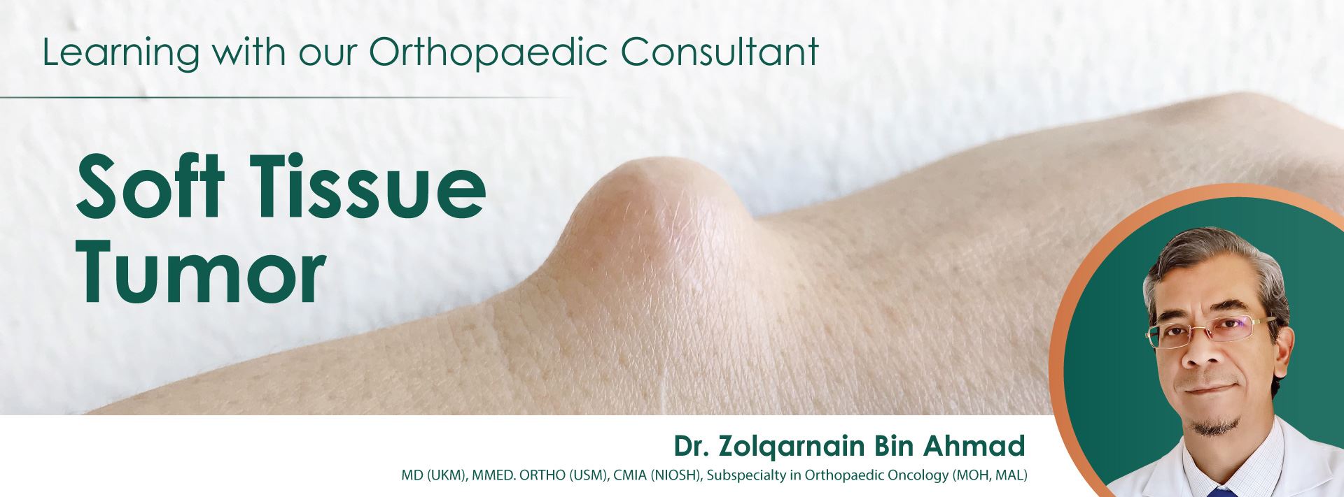
| STS is a growth from the muscle, fat, blood vessels, nerve, fibrous tissue etc. They can be found in any part of your body.
|
|
|
What is the common warning or alarming signs?
|
- The lump is large, according to the guidelines: > 5cm
- Swelling in deep layers
- It grows quickly
- Skin changes, such as blood vessel dilation, bleeding, rupture, ulceration, redness
- Disruption of limb function
|
Type of Soft Tissue Tumor (STS)
|
STS can be benign or malignant.
The benign form is usually slow growing, less harmful, does not spread or invade locally, and does not spread to distant organs.
Whereas the malignant type is cancerous, it spreads to local and distant organs such as the lungs and liver. Malignant types require urgent attention.
|
|
|
Disease Demographics
|
STS affects men and women with equal frequency. It is more common in adults and accounts for 10% in children.
Benign soft tissue tumors are 10 times more common than malignant tumors.
|
|
|
Causes, Risk Factors and Prevention
|
The causes of STS are mostly unknown and there are no known ways to prevent them.
Although there are no obvious risk factors, possible risk factors include radiation, family cancer syndromes, genetic and chemical factors.
|
|
|
Investigation
|
| MRI is an investigative modality that provides an accurate description of the tumor. It is mandatory for large and deep masses. Regional X-ray rules out bony involvement. For staging purposes, MRI and CT scans are mandatory and very important investigations. A biopsy is required to establish an accurate histopathological diagnosis.
|
|
|
Treatment
|
| STS treatment is individualized according to diagnosis, type, tumor origin, structural involvement, etc. Often, the most likely option is surgery.
|
|
|
Prognosis
|
Although benign tumors are not cancerous and do not spread or invade locally, they can cause serious problems, such as lesions compressing nerve pathways, blood vessels and interfering with limb function.
Malignant soft tissue tumors can be life-threatening.
|
|
|
How we at KGSC can help you?
|
- If you have a lump or swelling at any part of your body, please come to us for a consultation.
- If you have warning signs, you may need urgent proper, thorough investigation and treatment.
- Early detection, evaluation and treatment provide better outcomes and function.
Treatment Process
- Consultation: By appointment or walk in
- Investigation: Blood test, X-ray, Ultrasound, CT, MRI and Biopsy
- Definitive Treatment: Surgery
|
|
|
| My Personal Advice: Don't wait until the swelling is big because the surgery will become complicated, long incisions, hours of surgery, risk of bleeding or blood loss and affect the function of the limb after surgery.
|
|
|
| For more information or appointment, please call us at +607-2133899 or email info@kgsc.com.my.
|
|
|
|
|
| Kawasaki Disease
|
Dr. Choong Choun Seng
|
|
Kawasaki Disease
|
Kawasaki Disease, also known as Mucocutaneous Lymphnode Syndrome, was first reported by Japanese pediatrician Tomisaku Kawasaki in 1970 and was named after him. It is mainly systemic vascular inflammation. So far, the cause is unknown.
|
|
| This is an acute systemic vasculitis of childhood. If the age is less than 5 years old, the diagnosis is not timely and the golden treatment period is missed, coronary aneurysm may form. Once ruptured, it is likely to be life-threatening.
|
|
|
The Main Symptoms of Kawasaki Disease
|
|
- High fever above 38 degrees and last for more than 5 days
- Hyperemia in the whites of both eyes (Figure 1)
- Bleeding of oral mucosa and chapped lips (Figure 2)
- Strawberry tongue on the surface of the tongue (Figure 3)
- Red, swollen and peeling palms and feet (Figure 4)
- Unilateral nuchal lymphadenopathy
- Polymorphic rashes on the trunk
|
|
|
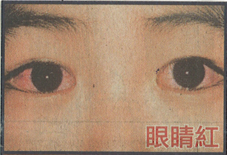
Figure 1
|
|
|
.jpg)
Figure 2
|
|
|
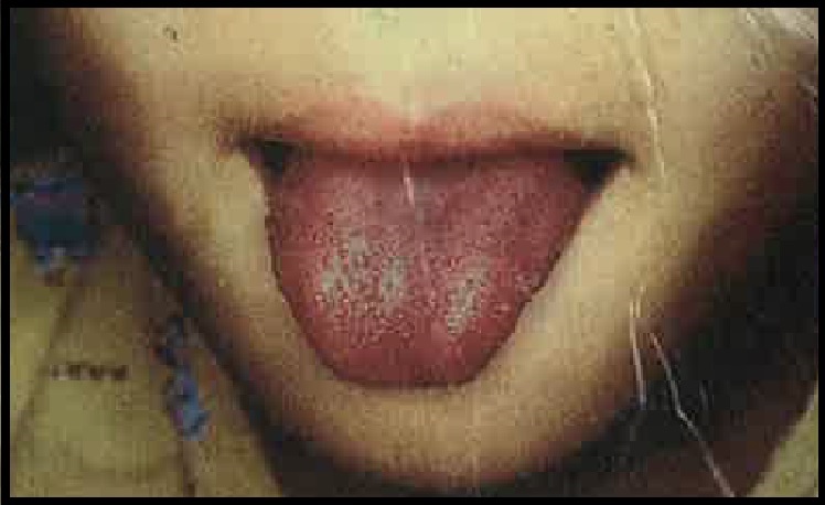
Figure 3
|
|
|
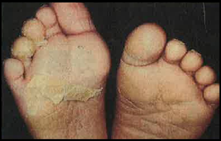
Figure 4
|
|
|
Treatment
|
| The treatment of Kawasaki disease should use high-dose immunoglobulin in time to avoid sequelae of coronary aneurysm. Rupture of a coronary aneurysm can lead to death if left untreated.
|
|
|
| Kawasaki disease often occurs in children under the age of 5, and the exact cause is still unknown. Many studies have found that it may be caused by a series of reactions in the body's immune system triggered by a special virus infection. These viruses include Adenovirus, Coronavirus, Enterovirus and Rhinovirus. Asian countries such as Japan, China, Taiwan, Hong Kong and South Korea all have many cases, as does Malaysia, but not many.
|
|
|
| At present, the medical community believes that Kawasaki disease is an abnormal human immune response that causes inflammation and damage to blood vessels. Large doses of immunoglobulin can suppress this immune response to control inflammation and treat diseases.
|
|
|
| To make an appointment with our paediatrician, please WhatsApp us at +6016-9535113 or call us at +607-2133899.
|
|
|
|
|
| Mycoplasma Pneumonia in Children
|
Dr. Choong Choun Seng
|
|
Mycoplasma Pneumonia in Children
|
Children's mycoplasma pneumonia is prone to occur in preschool children, especially kindergarten children.
In general, the pathogenic bacteria of community acquired pneumonia are some typical bacteria, including Streptococcus pneumoniae, Haemophilus influenzae, and Staphylococcus aureus (S. aureus). Symptoms are typically acute fever, chills (fever and chills), cough, rust-coloured phlegm, chest pain and shortness of breath.
|
|
| Atypical pneumonia is named for its mild symptoms and indistinct signs (sometimes flu-like symptoms such as chills, chills, fever, headache, obvious cough but less thick phlegm, loss of appetite). The most common pathogen is Mycoplasma pneumonia, followed by Chlymydophila pneumonia and Legionella pneumophila.
|
|
|
| The biggest difference between atypical pneumonia and typical bacterial pneumonia is that there is no toxic sign in atypical pneumonia, especially symptoms of shortness of breath (shortness of breath) and difficulty breathing (dyspnea), but a dry cough.
|
|
|
| Most cases of mycoplasma pneumonia progress slowly and usually manifest as pharyngitis, bronchitis, pneumonia, and otitis media. Common symptoms include dry cough, or paroxysmal severe cough (more obvious), rise in body temperature (fever lasts for 2-3 weeks), malaise, chills, headache, muscle or joint pain, sore throat, and ear pain. Usually, there is no runny nose and chest pain.
|
|
|
| Mycoplasma pneumonia is generally treated with erythromycin or tetracycline for 10-14 days. Most of the fever subsides quickly after treatment, and the uncomfortable symptoms are also relieved. Tetracyclines may harm developing bones and teeth, so they are not recommended for children under the age of eight.
|
|
|
Understanding Mycoplasma Pneumonia in One Simple Table
|
|
|
|
| Community Acquired Pneumonia
|
Pathogenic Bacteria
- Streptococcus pneumoniae
- Haemophilus influenzae
- Staphylococcus aureus (S. aureus)
|
|
|
Symptoms
- Chills, shivering
- Cough
- Rust-coloured phlegm
- Chest pain
- Shortness of breath
|
|
|
| Atypical Pneumonia
|
Pathogenic Bacteria
- Mycoplasma pneumonia
- Chlymydophila pneumonia
- Legionella pneumophila
|
|
|
Symptoms
- Mainly dry cough
- Loss of appetite
- There is no shortness of breath and difficulty in breathing
|
|
|
Treatment: Antibiotics
1. Erythromycin or Tetracycline
2. Duration of 10 - 14 days
3. Tetracycline should not be given to developing children ( ≤8 years old ), as it will affect the development of bones and teeth.
|
|
|
|
|
| To book an appointment with our paediatrician, please WhatsApp us at +6016-9535113 or call us at +607-2133899.
|
| Click here to request an appointment booking through our website.
|
|
|
|
|
|
|
|
|
|
|
Breast Self-Examination (BSE)
|

|
|
|
Breast Self-Examination (BSE)
|
|
|
| Breast self-examination is an easy way and can be a useful tool in detecting for breast cancer early when it is most treatable (Early detection is the best defence against breast cancer).
|
|
|
- The way breasts look, and feel is different for every woman. The goal of BSE is to become familiar with the way your breasts look and feel to you. Then, if there are any changes you are more likely to notice it earlier, so you can see your doctor as soon as possible.
- Examine your breasts once a month when they are least tender (usually 5-10 days from the first day of your period).
- If you no longer have periods, choose one fixed day each month that will remind you to do BSE.
- If you are breast feeding, empty your breasts first.
- Call your doctor or nurse if there are any changes.
- Remember, most breast changes are NOT cancer, but DO check up to be sure!
|
|
|
7P of Breast Self-Examination (BSE)
|
|
|
|
|
|
|
Position
|
|
|
|
|
| Standing
|
|
|
| Stand to look at your breasts: Standing undressed to the waist in front of a mirror in three positions:
|
- Arm relaxed at sides.
- Press your hands firmly on your waist and lean slightly towards the mirror.
- Arms raised above head.
|
|
|
| Look at your breasts for any changes in size, shape of the breasts, colour, texture of the nipples, skin and direction of your nipples point (notice if there is any retraction of the nipple). Observe any stain on your night clothes or bra from your nipples, especially if only from one side. If there is any change or look unusual, seek medical advice immediately.
|
|
|

|
|
|
| Lying Down
|
|
|
- Lie down on your back, so the breast tissue spreads more evenly over the chest wall, making it easier to feel all the breast tissue.
- Place your right arm behind your head. Placing a pillow or rolled towel under the right shoulder (under the breast from the back) may be helpful.
- Use the finger pads of the 3 middle fingers of your left hand to feel for lumps in the right breast. Do the same way for the left breast.
|
|
Perimeter (Where to Feel)
|

|
|
|
| The area to be examined should include all the breast tissue and the armpit (as shown on the picture).
|
|
|
Palpation with Pads of Fingers (How to Feel)
|
- Use over tapping dime-sized circular motions of your three middle finger pads to feel the breast tissue.
- Feel a small portion of the breast at a time until the entire breast has been checked.
- Do not lift your fingers from your breast between palpations. You can use powder or lotion to help your fingers slide from one spot to the next.
|
|
Pressure (How Deep to Feel)
|
- Use three levels of pressure for each palpation, from light to deep, to examine the full thickness of your breast tissue.
- Using pressure is important because the breast is not flat. You need to feel all the way through the tissue to your ribs.
|
|
Pattern of Search
|
|
|
|
|
- Lines: Beginning at the outer edge of your breast, move your fingers downward using a circular motion until they are below the breast. Then move your fingers slightly toward the middle and slowly move back up.
- Circles: Beginning at the outer edge of your breast, use the flat part of your fingers, moving in circles slowly around the breast. Gradually make smaller and smaller circles toward the nipple. Be sure to check behind the nipple.
- Wedges: Starting at the outer edge of the breast, move your fingers toward the nipple and back to the edge.
|
|
Practice with Feedback
|
- Have your doctor or nurse show you how to do BSE.
- Then ask them to watch you do the exam to see that you are doing it right.
|
|
Plan of Action
|
| You should have a personal breast health plan:
|
- Discuss breast cancer early detection guidelines with your doctor or nurse.
- Schedule your clinical breast exam and mammogram as appropriate for your age.
- Perform BSE monthly.
- Report any breast changes to your doctor or nurse.
|
|
|
|